
What does this procedure involve?
This involves using a telescope (semi-rigid or flexible) passed into your bladder through your urethra (waterpipe) to fragment and/or remove stones from your ureter or kidney. We sometimes need to leave a temporary stent in your ureter after the procedure.
What are the alternatives?
- Observation – most stones smaller than 5mm in diameter pass by themselves but larger stones (greater than 7mm diameter) rarely do
- Extracorporeal shockwave lithotripsy (ESWL) – this involves using a machine that generates shock waves which travel through your skin to break the stone into fragments which you then pass yourself
- Antegrade ureteroscopy – stones in the upper ureter (close to the kidney) can be treated by percutaneous (keyhole) puncture of the kidney so that a ureteroscope can be passed down to the stone from above
- Laparoscopic (keyhole) or robotic stone removal – for extremely large or impacted stones in the ureter
- Percutaneous nephrolithotomy (PCNL) – for large stones in the kidney which may not be suitable for flexible ureteroscopy
- Open stone removal – although very unusual nowadays, if all the above techniques fail we may need to resort to open surgery, through an incision in your side, to remove your stone(s)
What happens on the day of the procedure?
Your urologist (or a member of their team) will briefly review your history and medications, and will discuss the surgery again with you to confirm your consent.
An anaesthetist will see you to discuss the options of a general anaesthetic or spinal anaesthetic. The anaesthetist will also discuss pain relief after the procedure with you.
We may provide you with a pair of TED stockings to wear, and we may give you a heparin injection to thin your blood. These help to prevent blood clots from developing and passing into your lungs. Your medical team will decide whether you need to continue these after you go home.
If you have a stone in your ureter, we usually arrange an X-ray or, occasionally, a CT scan for you on the day of the operation, to be sure that the stone has not passed by itself.
Details of the procedure
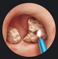
- we normally use a full general anaesthetic and you will be asleep throughout the procedure
- we usually give you an injection of antibiotics before the procedure, after you have been checked for any allergies
- we put a telescope into your bladder, through your urethra (waterpipe), and use it to put a guidewire up into your kidney, past the stone in the ureter, using X-ray control
- we then put the ureteroscope (semi-rigid or flexible) up to the level of the stone by following the guidewire
- we fragment the stone using an energy source; this is usually a laser but, occasionally, we use a lithoclast (a tiny pneumatic drill)
- using the laser, we either “dust” the stone, leaving tiny fragments which can pass by themselves, or break it into smaller pieces (pictured) which can be removed using special grasping devices
- we may insert a temporary drainage tube (a ureteric catheter, or a stent with a string attached) into the ureter at the end of the procedure; this is removed later and your urologist will arrange this for you
- occasionally, we need to perform a “second-look” ureteroscopy at a later stage to treat residual stones; if this is needed, we leave a stent in your ureter until the second procedure
- occasionally, we put in a bladder catheter which is removed the following morning
- many patients will be able to go home on the same day as their procedure or early on the first post-operative morning
If you have been admitted as an emergency (with a stone blocking your ureter), you may have had several tests and other treatment already. This means your stay in hospital will, inevitably, be longer than a single day.
Further information and a short video of ureteroscopic stone removal are available on the BAUS website.
Are there any after-effects?
The possible after-effects and your risk of getting them are shown below. Some are self-limiting or reversible, but others are not. The impact of after-effects can vary a lot from patient to patient; you should ask your surgeon’s advice about the risks and their impact on you as an individual:
| After-effect | Risk |
|---|---|
| Mild burning or bleeding on passing urine for a short time after the procedure (especially if you have a ureteric stent) |
 Almost all patients Almost all patients |
| Temporary insertion of a ureteric stent which needs to be removed later |
Almost all patients |
| Recurrent (new) stone formation over the next five to 10 years, requiring further surgery or other treatment |
 1 in 2 patients (50%) 1 in 2 patients (50%) |
| Residual stones requiring further surgery or other treatment (more likely for stones closer to the kidney) |
 Between 1 in 7 & 1 in 20 patients (5 to 15%) Between 1 in 7 & 1 in 20 patients (5 to 15%) |
| Failed to access the ureter (or reach the stone) requiring further surgery or other treatment |
1 in 20 patients (5%) |
| Temporary insertion of a bladder catheter |
Between 1 in 10 & 1 in 50 patients |
| Infection requiring antibiotic treatment |
Between 1 in 50 & 1 in 100 patients |
| Minor damage to the wall of the ureter (small perforation, mucosal abrasion, bleeding) requiring stenting or percutaneous nephrostomy |
 1 in 100 patients (1%) 1 in 100 patients (1%) |
| Narrowing of the ureter due to delayed scar formation (stricture) which may require further treatment |
Between 1 in 100 & 1 in 250 patients |
| Major damage to the wall of the ureter (large perforation, avulsion of the ureter) requiring further surgery |
Less than 1 in 1000 patients (less than 0.1%) |
| Anaesthetic or cardiovascular problems possibly requiring intensive care (including chest infection, pulmonary embolus, stroke, deep vein thrombosis, heart attack and death) |
Between 1 in 50 & 1 in 250 patients (your anaesthetist can estimate your individual risk) |
What is my risk of a hospital-acquired infection?
Your risk of getting an infection in hospital is between 4 & 6%; this includes getting MRSA or a Clostridium difficile bowel infection. This figure is higher if you are in a “high-risk” group of patients such as patients who have had:
- long-term drainage tubes (e.g. catheters);
- bladder removal;
- long hospital stays; or
- multiple hospital admissions.
What can I expect when I get home?
- you will be given advice about your recovery at home
- you will be given a copy of your discharge summary and a copy will also be sent to your GP
- any antibiotics or other tablets you may need will be arranged & dispensed from the hospital pharmacy
- you should drink twice as much fluid as you would normally for the first 24 to 48 hours, to flush your system through and reduce the risk of infection
- recovery from ureteroscopy is usually rapid; you may return to work when you are comfortable enough and when your GP is satisfied with your progress
- if you have had a stent put in, it may cause pain in your kidney area when you pass urine, or pain in your bladder; this usually settles quickly but, if you feel unwell or feverish, you should contact your GP to check for a urine infection
- if you develop a fever, pain in the area of the affected kidney, severe pain on passing urine, inability to pass urine or worsening bleeding, you should contact your GP immediately
You can reduce your risk of further stone formation by altering your diet and fluid intake. Ask your urologist or specialist nurse for further details about this or download the BAUS leaflet “Dietary advice for stone formers”.
General information about surgical procedures
Before your procedure
Please tell a member of the medical team if you have:
- an implanted foreign body (stent, joint replacement, pacemaker, heart valve, blood vessel graft);
- a regular prescription for a blood thinning agent (e.g. warfarin, aspirin, clopidogrel, rivaroxaban, dabigatran);
- a present or previous MRSA infection; or
- a high risk of variant-CJD (e.g. if you have had a corneal transplant, a neurosurgical dural transplant or human growth hormone treatment).
Questions you may wish to ask
If you wish to learn more about what will happen, you can find a list of suggested questions called “Having An Operation” on the website of the Royal College of Surgeons of England. You may also wish to ask your surgeon for his/her personal results and experience with this procedure.
Before you go home
We will tell you how the procedure went and you should:
- make sure you understand what has been done;
- ask the surgeon if everything went as planned;
- let the staff know if you have any discomfort;
- ask what you can (and cannot) do at home;
- make sure you know what happens next; and
- ask when you can return to normal activities.
We will give you advice about what to look out for when you get home. Your surgeon or nurse will also give you details of who to contact, and how to contact them, in the event of problems.
Smoking and surgery
Ideally, we would prefer you to stop smoking before any procedure. Smoking can worsen some urological conditions and makes complications more likely after surgery. For advice on stopping, you can:
- contact your GP;
- access your local NHS Smoking Help Online; or
- ring the free NHS Smoking Helpline on 0300 123 1044.
Driving after surgery
It is your responsibility to make sure you are fit to drive after any surgical procedure. You only need to contact the DVLA if your ability to drive is likely to be affected for more than three months. If it is, you should check with your insurance company before driving again.